What is the Histology Atlas?

The BEST Network Histology Atlas is a comprehensive resource that covers the study of microscopic structures of normal tissues and cells. It complements the virtual slides available on Slice and provides academics with a curated, ready-to-use resource to help them get started building a new histology course or supplement an existing course.Students can use the annotated images and explanations to support their study of histology or as a reference resource while studying histopathology.

Try demo
Chest xraySlice utility bar

Self-paced online resource

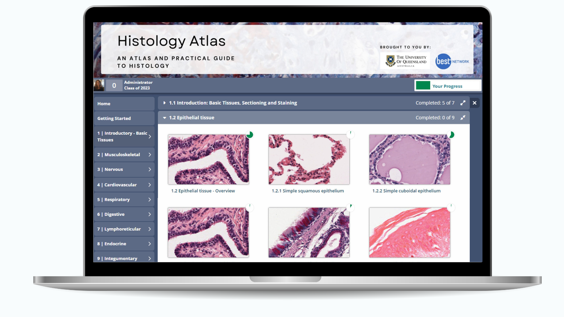
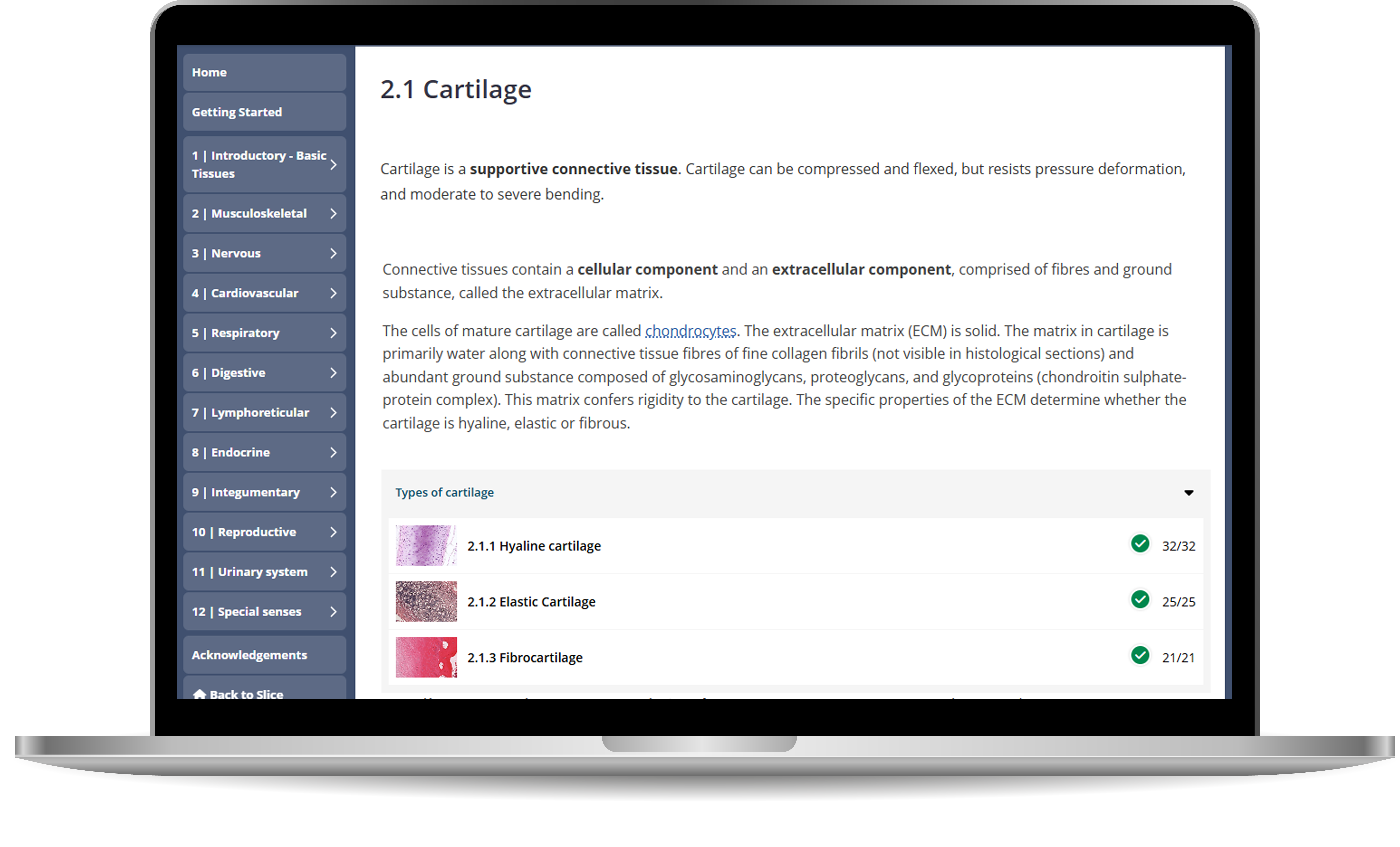
Built within the award winning OpenLearning platform, our Atlas offers a self-paced approach to learning histology.Organised by body system, students can use it to review any tissue or organ and can complete as much or as little as they need. Search tools can help students immediately find what they are looking for.

Try demo

Detailed histological study

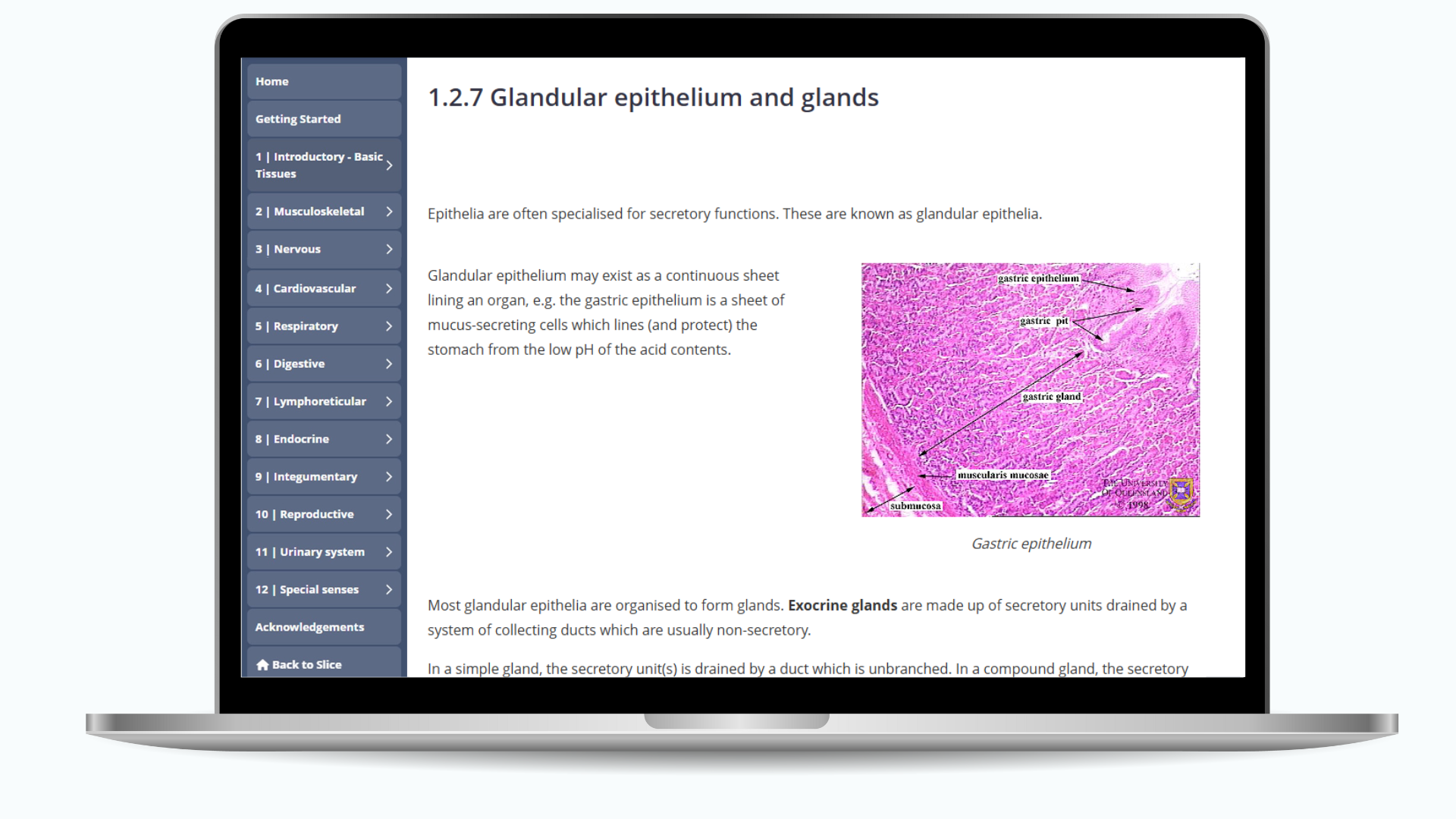
Pages focus on function and histological appearance and include text-based descriptions, combined with annotated virtual slides, static labelled images and diagrams to ensure learners receive a well-organised and in-depth education on each subject.

Learn more

Annotated Layers

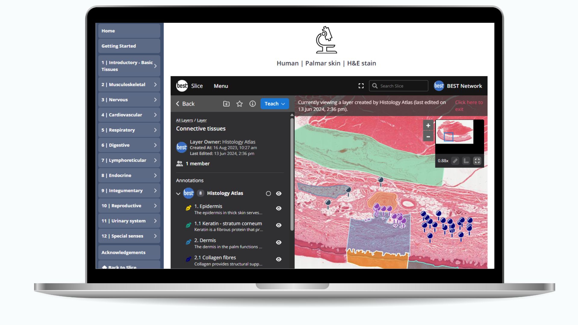
Dive deeper into histological structures with annotated layers that provide insightful context and explanations. Annotations support students in their examination of features and virtual slides allow them to be viewed at any magnification.Wherever possible multiple examples of features have been marked to help students recognise normal variation.

Try demo
Slice deep cell micro imageutility bar

Test your knowledge

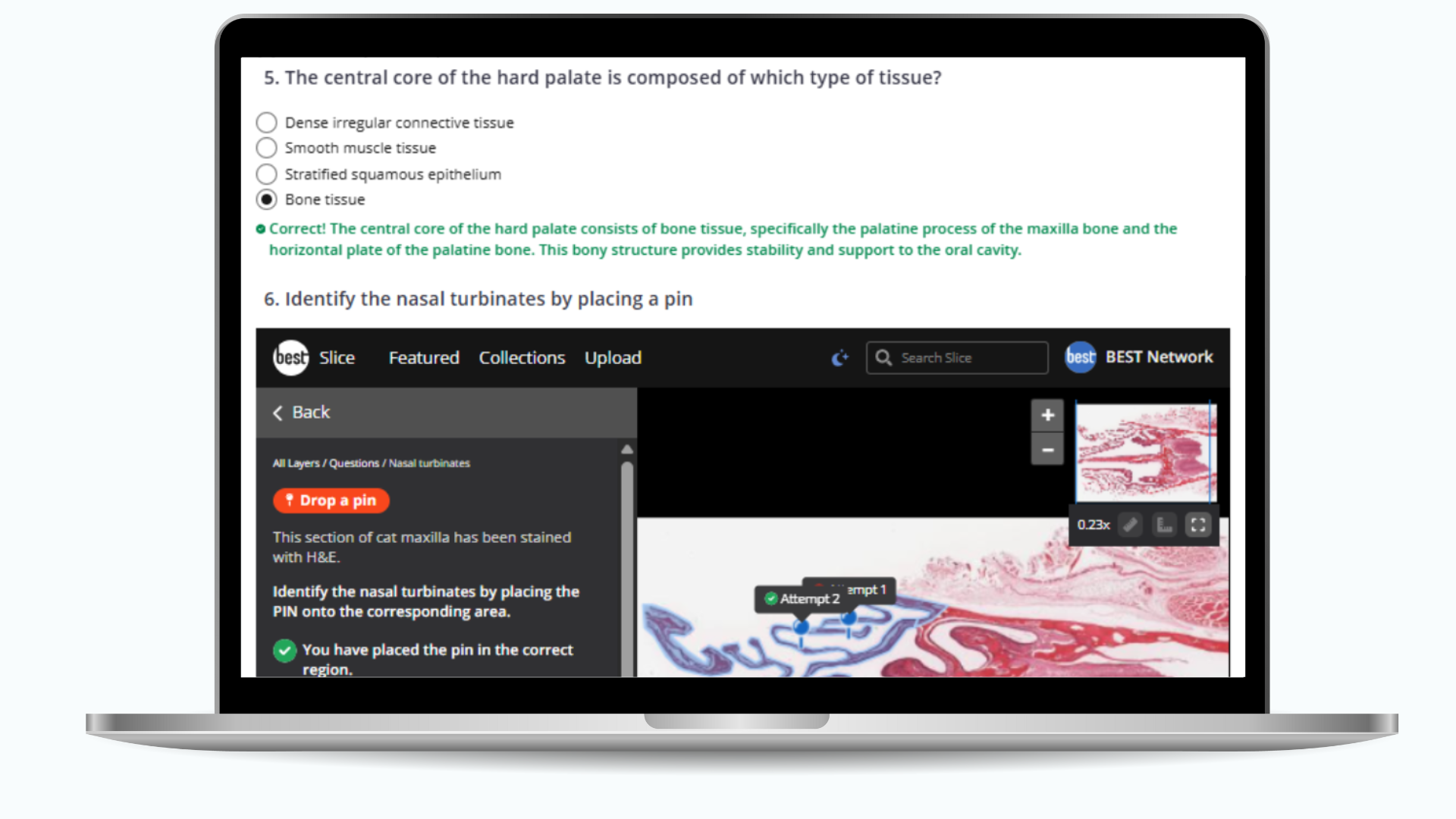
End of topic quiz questions allow students to test their knowledge and receive immediate feedback on function and histological appearance using a variety of multiple choice questions, matching and sorting questions and ‘Drop a Pin’ questions that examine feature identification skills.

Try demo
testimonials
"The newly developed online modules (SLICE) in pathology were beneficial for self-study and for applying our pathology lecture and practical knowledge. Please continue making some of these amazing tools"
Second Year Medical Student
Griffith University, 2016
"All pathology pracs are around virtual slides in Slice, and our online tutorials are around macroscopic specimens in Slice, so Slice is essential to our online teaching."
Professor Martin Weber
UNSW Sydney, 2021
"Slice helps me identify if students are able to apply theory content that we have covered to real slides."
Dr Noelia Roman
Charles Sturt University, 2021
"I was able to use my student Slice account for the first time in a med histology prac yesterday. I thought it was fantastic. I don’t think I’ve learnt so much in a histology lab before, cause now it’s just so easy to take notes while still keeping up with where the lecturer is on the slide."
Medical Student
UNSW Sydney
"The Slice system has been a godsend for teaching during COVID-19"
Professor Shane Raidal
Charles Sturt University, 2022
"My whole pathology teaching is based on Slice"
Dr Suja Pillai
University of Queensland, 2022
"This is the first time biomedical experts have come together to create a common database of complex, rare and exceptionally valuable educational resources of the highest quality that are shareable among different institutes.  BEST is a valuable tool in improving health care education in Australia, with potential for global utility.”
Professor Shashidar Venkatesh, Head of Pathology
James Cook University

3,599

Anatomy (Human)

7,915

Pathology (Human)

2,474

Veterinary Science

2,291

Diagnostic Imaging

376

Microbiology

577

Haematology

259

Botany

396

Dentistry

Zoom and pan viewing

View all images at higher magnification. Navigate virtual slides just as you would under the microscope, zooming in/out up to 40x-100x and panning across the tissue section.

Try demo
Slice engagement UISlice utility barAnnotation pinAnnotation pin

Engaging Group Activities

Invite students to work together on a shared layer and evaluate their annotations.

Try demo
Slice deep cell micro imageutility bar

Immersive Presentations

Remove distractions and focus on explaining the features of an image to students.

Try demo

Integrate the Histology Atlas into your class activities

  • Each page has a unique URL that can be used to recommend reading and preparation for your students before class.
  • The atlas provides a comprehensive starting place for those looking to create a new histology course, with over 350 annotated images.
  • Ideal for both histology and histopathology to help students quickly revisit the histological appearance of normal tissue.

We believe that the Histology Atlas will be an invaluable addition to your teaching toolkit, making your job easier while enhancing the educational experience for your students.

Get Started

To get started with the Histology Atlas and explore its full potential, simply log in to your Slice account and navigate to the new "Histology Atlas" section. It is included free with your existing membership to Slice!

Get started

To get started with the Histology Atlas and explore its full potential, simply log in to your Slice account and navigate to the new "Histology Atlas" section. It is included free with your existing membership to Slice!

Join now